The Premier Continuing Education
Provider |
|||
|
|
Massage Awareness - The Standard For Animal Massage Continual Education |
|||
| Thermal Imaging | |||
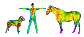
Use Infrared Imaging for Injury Identification & Prevention Stop Guessing… Start Knowing! Thermal imaging maps the heat signature of your animal allowing you to see what you couldn’t before. Animals by nature mask their pain but with a thermal imager we are able to see the hot spots point blank. |
|||
What is Thermal Imaging Thermal Imaging is a non-invasive, completely safe technology that has the ability to detect changes in temperature related to localized inflammation and/or blood flow to the tissues. Such changes have been shown to occur 2-6 weeks prior to the onset of clinical signs of lameness. Infrared thermography is a diagnostic tool that uses the very latest Infrared imaging equipment and computer software to detect minute differences in the horse's thermal and neural condition and allows us to quickly and efficiently identify trauma in an injured animal. By identifying the location of the injury we can prevent further trauma, make a decision on treatment needed and monitor the recovery. Infra red thermal imaging of the Equine has proved to be a valuable tool in the diagnosis and prognosis of the injured Equine. What do you use it for? Thermal imaging is a physiologic imaging modality, therefore it detects changes in blood flow and metabolism, but it cannot necessarily tell you which exact anatomic structure is affected. Thermal imaging detects surface heat directly correlated to circulation, or a lack thereof. Therefore, anything that requires a tool to identify inflammation, reduced circulation, potential nerve damage, or serial patterns, falls under the thermal imaging umbrella. How does it works? Infrared thermography identifies abnormalities by measuring the surface temperature of an animal’s body and legs, showing areas that are excessively warm or cool, both of which may reveal an abnormality indicative of a problem. Thermal Imaging can detect changes with a thermal sensitivity of 0.04 degree Celsius. This allows you to perceive early abnormalities before they become anatomical disruption such as muscle strain, ligament sprain, joint capsule inflammation, stress fractures and more. If not detected early some condition may become catastrophic or even life-threatening situations. Equipment We use the FLIR T300 camera for all our consultations because we believe it to be the best equipment in the world today, enabling the capture of the highest resolution infrared images possible and maximizing the contrast optimization options. Combined with advanced software (FLIR reporter 8.5 Pro Software), we can provide you a detailed report with photographical support in either a printed form or as a pdf file that can be emailed to other professional at your discretion. Thermal Imaging versus X-ray and MRI Unlike conventional X-rays and MRI scans, Infrared Thermographic Imaging does not use any radiation and is therefore perfectly safe for the animal and the handler. The only other physiologic imaging modality is nuclear scintigraphy, or a “bone scan,” which involves injecting a radiopaque isotope that highlights areas of inflammation. Anatomic imaging modalities include traditional radiographs (“x-rays”), ultrasound, Computed Tomography (CT) and Magnetic Resonance Imaging (MRI). These modalities pinpoint the structures affected with pathology, but only give a static image of disease processes. Physiological imaging such as infrared thermal Imaging has the ability to not only see tissue conditions but can also recognize areas that are indicative to anatomical tissues. In most cases the results of Thermographs can be provided instantly to enable vets, owners and animal therapists and professionals to make prompt diagnoses and begin appropriate treatment.
|
Take away the “Guessing” – Uses of Thermal Imaging Early detection is your best Prevention
Common Uses of Thermal Imaging
Benefits
*Travel Fee may apply. Submit your request via our website for complete pricing in your area |
||
Home | About Us | JP Bio | Contact Us | CEUs | FAQ | Certification Program | Courses | Practical Training | Registration | Classes & Fees | Resources | What's New | Testimonials | Site Map | Upcoming Courses | Online Exam | NCBTMB | IAAMB/ACWT | FRENCH Animal Massage Awareness Store | Join Our Mailing List    | |||
| | |||
| © 2021 Massage Awareness, Inc. - Site Design by Carlton Design Concepts | |||